How high blood pressure kills you
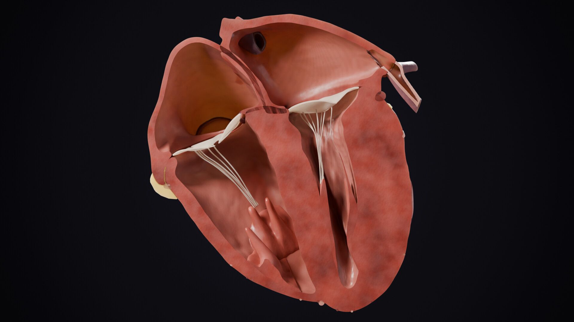
High blood pressure thickens the heart until it struggles to pump, leaves small strokes in the brain that go unnoticed, scars the kidneys, and can split the largest artery from the inside. Most people feel nothing while the damage builds. Across more than a hundred trials, lowering the top number by ten points cut strokes by about a quarter.
Claims and sources
- 01
Nearly half of American adults have it, and most feel nothing.
On screen
47.7 %
US adults · NHANES 2021–23
The survey counts adults whose measured pressure was above its cut-offs or who were taking medication for it; other bodies draw the line elsewhere, and the film gives no number for what counts as high. “Most feel nothing” is the WHO’s wording. The same page says very high pressures can cause headaches, blurred vision and chest pain.
During August 2021–August 2023, the prevalence of adult hypertension was 47.7%.
Fryar CD, Kit B, Carroll MD, Afful J. Hypertension prevalence, awareness, treatment, and control among adults age 18 and older: United States, August 2021–August 2023. NCHS Data Brief No. 511, October 2024 (NHANES).
Most people with hypertension don't feel any symptoms.
World Health Organization. Hypertension (fact sheet), 25 September 2025.
- 02
Start with your smallest arteries. They are narrow and muscular, and they set about 70 percent of the resistance your heart pushes against.
On screen
about 70 %
Share of resistance · small arteries and arterioles
The source says “in most organs”; the film says what the heart pushes against. “Narrow and muscular” is description, not a sentence from this source.
As a group, changes in diameter (and therefore resistance) of small arteries and arterioles have the greatest influence on vascular resistance because these two vessel segments comprise about 70% of the total resistance in most organs.
Klabunde RE. Cardiovascular Physiology Concepts (online edition of the textbook, cvphysiology.com).
- 03
And resistance is brutal about width. Narrow one of them by ten percent, and its resistance jumps about fifty percent.
On screen
+52 %
Resistance · radius −10 % · idealised flow, one artery
Our arithmetic, not a figure from the source: resistance varies as the inverse fourth power of the radius, so 1 divided by 0.9 to the fourth is about 1.52. It is one artery in an idealised law of flow; real vessels differ.
Resistance is inversely proportional to the radius to the fourth power.
inversely proportional to the radius to the fourth power (r4) or diameter to the fourth power (D4).
Klabunde RE. Cardiovascular Physiology Concepts (online edition of the textbook, cvphysiology.com).
- 04
Under high pressure their walls thicken, and the opening shrinks. Narrower means more resistance. More resistance means more pressure. The loop feeds itself.
The sources describe the thickening and the smaller opening; the loop is our summary of the two together.
In chronic hypertension, vessel radius is often reduced because of a thickening of the vessel wall - this leads to a reduction in lumen size.
Klabunde RE. Cardiovascular Physiology Concepts (online edition of the textbook, cvphysiology.com).
This increased resistance is caused by a thickening of the walls of resistance vessels and by a reduction in lumen diameters.
Klabunde RE. Cardiovascular Physiology Concepts (online edition of the textbook, cvphysiology.com).
- 05
Your big arteries should stretch with every beat and soften the pulse. Under pressure they stiffen, and each pulse lands harder on your brain and kidneys. High pressure can also damage the lining of arteries, helping cholesterol plaques grow.
The kidney and brain sentence comes from a review of ageing and aortic stiffness; it does not name high blood pressure as the cause of the stiffening (the first source does). The plaque sentences are about the artery wall and its permeability to cholesterol, and describe a contribution, not the whole cause.
The excessive collagen can replace the more elastic fibers, making the arteries more rigid and less able to expand and recoil with blood flow.
Kim HL. Arterial stiffness and hypertension. Clin Hypertens. 2023;29:31. PMID 38037153. (Source given by the owner, 2026-11-12.)
When BP is high, arterial stiffness progresses, and conversely, when arterial stiffness progresses, BP also increases.
Kim HL. Arterial stiffness and hypertension. Clin Hypertens. 2023;29:31. PMID 38037153. (Source given by the owner, 2026-11-12.)
In high-flow (low-impedance) organs, like the brain and kidneys, pressure pulsatility penetrates further into the microcirculation.
Mitchell GF. Effects of central arterial aging on the structure and function of the peripheral vasculature: implications for end-organ damage. J Appl Physiol 2008;105(5):1652-1660.
Over time, high blood pressure can damage artery walls, allowing plaque to build up.
NHLBI. Atherosclerosis: Causes and Risk Factors (last updated October 28, 2024).
In hypertension, dysfunction of the artery’s inner lining may be involved in vascular inflammation, remodeling and atherosclerosis.
Gallo G, Volpe M, Savoia C. Endothelial dysfunction in hypertension: current concepts and clinical implications. Front Med 2022;8:798958.
Hypertension may increase the artery wall’s permeability to LDL cholesterol, raising the risk of accelerated atherosclerosis.
Nielsen LB. Transfer of low density lipoprotein into the arterial wall and risk of atherosclerosis. Atherosclerosis 1996;123(1-2):1-15.
- 06
Your heart pushes against all of this, about a hundred thousand times a day. So it does what muscle does. It gets thicker. But a thick wall is a stiff wall. It takes more pressure to fill. That pressure backs up into the lungs. Stairs leave you breathless.
On screen
≈ 100,000 beats a day
Our arithmetic from a resting range of 60 to 100 a minute
The daily count is arithmetic: 60 to 100 beats a minute is 86,400 to 144,000 a day, and 100,000 a day is about 69 a minute. The stiff-wall passage describes the chronic pressure-overloaded heart; breathlessness on stairs is the source’s example of an early symptom.
A resting pulse of 60 to 100 beats per minute is the listed normal range for children 10 and older and adults.
MedlinePlus Medical Encyclopedia. Pulse (reviewed January 1, 2025).
With chronic pressure overload, as occurs with chronic hypertension or aortic valve stenosis, the ventricular chamber radius may not change; however, the wall thickness increases as new sarcomeres are added in-parallel to existing sarcomeres.
Klabunde RE. Cardiovascular Physiology Concepts (online edition of the textbook, cvphysiology.com).
This type of ventricle becomes "stiff" (i.e., compliance is reduced), which can impair filling and lead to diastolic dysfunction.
Klabunde RE. Cardiovascular Physiology Concepts (online edition of the textbook, cvphysiology.com).
The elevated left ventricular filling pressure is transmitted back into the left atrium and pulmonary vasculature, thereby increasing left atrial pressure (LAP), pulmonary venous pressure (PVP), and pulmonary arterial pressure (PAP).
Klabunde RE. Cardiovascular Physiology Concepts (online edition of the textbook, cvphysiology.com).
One of the first symptoms you may notice is feeling short of breath after routine activities like climbing stairs.
NHLBI. Heart Failure: Symptoms (last updated March 24, 2022).
- 07
That is heart failure. The strange part: the squeeze of the heart can still look normal. It still pumps about the same amount at rest. But it takes high pressure to fill, and it can’t keep up when you move.
The picture is schematic: no output or ejection-fraction value is shown. The textbook hedges (“changes very little”), and “about the same amount at rest” is output over time, which rests on the review below. This is one of two kinds of heart failure the film shows; the other follows a heart attack.
Diastolic dysfunction and HF with preserved ejection fraction are the most common cardiac complications of hypertension.
Messerli FH, Rimoldi SF, Bangalore S. The transition from hypertension to heart failure: contemporary update. JACC Heart Fail. 2017;5(8):543-551. PMID 28711447.
A stiff ventricle leads to reduced filling (decreased end-diastolic volume, EDV) and elevated end-diastolic pressure (EDP).
as klabunde-cvpc. Page hf006, 'Ventricular Diastolic Dysfunction (HFpEF)', revised 01/24/2024.
Cardiac output is typically normal at rest in patients with HFpEF, but the ability to increase cardiac output with exertion is frequently abnormal (33; 72), leading to impaired aerobic capacity.
Omote K, Verbrugge FH, Borlaug BA. Heart failure with preserved ejection fraction: mechanisms and treatment strategies. Annu Rev Med. 2022;73:321-337. PMID 34379445.
- 08
The heart’s own arteries collect plaque. When a plaque breaks open and a clot blocks a coronary artery, that is a heart attack. A heart attack kills muscle. What’s left can stretch and weaken, until the heart struggles to pump.
High blood pressure’s link to heart attack here is an association from a case-control study; it says nothing about plaque. No source says the dead muscle is “scarred”, so the film says dead muscle. The stretching of the surviving wall is from a review of remodeling after a heart attack.
Eventually, an area of plaque can break open inside your artery.
NHLBI. Heart Attack: Causes and Risk Factors (last updated March 24, 2022).
This causes a blood clot to form on the plaque's surface.
NHLBI. Heart Attack: Causes and Risk Factors (last updated March 24, 2022).
history of hypertension (1.91, PAR 17.9%)
Yusuf S, Hawken S, Ounpuu S, et al.; INTERHEART Study Investigators. Effect of potentially modifiable risk factors associated with myocardial infarction in 52 countries (the INTERHEART study): case-control study. Lancet. 2004;364(9438):937-952. PMID 15364185.
The noninfarcted region also undergoes an important lengthening that is consistent with a secondary volume-overload hypertrophy and that can be progressive.
Pfeffer MA, Braunwald E. Ventricular remodeling after myocardial infarction. Experimental observations and clinical implications. Circulation. 1990;81(4):1161-1172.
In heart failure with reduced ejection fraction (HFrEF), the left side of your heart is weak and can't pump enough blood to the rest of your body.
National Heart, Lung, and Blood Institute. Heart Failure: Causes and Risk Factors.
- 09
Backed-up pressure also stretches the chamber above, and its rhythm can turn chaotic. Clots can form there and travel to the brain.
The first source is about the high-pressure, thick-walled heart; the film draws the same back-up on the stretched heart after a heart attack. The clot source puts most clots in the chamber’s pouch (the appendage) in atrial fibrillation. “Can turn chaotic” is the film’s word for atrial fibrillation.
In addition to eliciting left ventricular hypertrophy, dysfunction and heart failure, hypertension also causes left atrial remodeling that may culminate in atrial contractile dysfunction and atrial fibrillation.
Kockskämper J, Pluteanu F. Left atrial myocardium in arterial hypertension. Cells 2022;11(19):3157.
Clots that break off and travel to the brain can cause a stroke.
MedlinePlus Medical Encyclopedia (National Library of Medicine). Atrial fibrillation or flutter (review date 1/1/2025).
201 of 222 clots in the left atrium in non-rheumatic atrial fibrillation were in, or began in, its pouch (the appendage).
Blackshear JL, Odell JA. Appendage obliteration to reduce stroke in cardiac surgical patients with atrial fibrillation. Ann Thorac Surg. 1996;61(2):755-759. PMID 8572814.
- 10
The brain has its own weak spot, deep inside. Tiny arteries branch straight off a large, fast one, and take its force. Over years their walls thicken and turn glassy. Some close. Some burst.
The first sentence is from the introduction of an imaging study and cites other work; no branching angle is claimed. The review speaks of reduced flow and small infarcts, not literally closure; the older review gives occlusion of a small vessel as one cause. “Glassy” translates hyaline. The picture is a diagram on a real brain scan, not a vessel-by-vessel map.
This abrupt size and flow change make LSAs especially susceptible to damage (e.g., hypertension).
Xu X, Wu X, Zhu C, et al. Characterization of lenticulostriate arteries and its associations with vascular risk factors in community-dwelling elderly. Front Aging Neurosci 2021;13:685571.
The pathologies thought to be secondary to uncontrolled hypertension include hyaline arteriolosclerosis (figure 1A), hyperplastic arteriolosclerosis, segmental arterial disorganization, and microaneurysm (figure 1B).
Cannistraro RJ, Badi M, Eidelman BH, Dickson DW, Middlebrooks EH, Meschia JF. CNS small vessel disease: a clinical review. Neurology 2019;92(24):1146-1156.
rupture results in hypertensive cerebral hemorrhage, typically in the subcortical white matter and deep gray nuclei.
Cannistraro RJ, Badi M, Eidelman BH, Dickson DW, Middlebrooks EH, Meschia JF. CNS small vessel disease: a clinical review. Neurology 2019;92(24):1146-1156.
Small lacunes are usually due to lipohyalinosis, larger ones to atheromatous or embolic occlusion of a penetrating vessel.
Fisher CM. Lacunar strokes and infarcts: a review. Neurology. 1982;32(8):871-876.
- 11
Either way, it’s a stroke. High blood pressure is tied to about half of all strokes.
On screen
≈ 48 %
Of strokes · population-attributable · INTERSTROKE
“Tied to”, not “caused by”: a case-control study of first strokes in 32 countries and 26,919 people, which counts a history of high blood pressure or a measured high reading. The 48 percent is the population-attributable risk (47.9 percent).
Hypertension was associated with all stroke (odds ratio 2.98), with a population-attributable risk of 47.9 percent.
O'Donnell et al. (INTERSTROKE). Lancet 2016;388(10046):761-75. PMID 27431356. 32 countries, 26,919 people.
- 12
Many small ones go unnoticed. Scans find them in up to one in five older adults with no symptoms. Unnoticed is not harmless. As damage adds up, it can slow walking, upset balance or dull thinking.
On screen
up to 1 in 5
Older adults · no symptoms · small deep strokes on MRI
The one-in-five sentence has no reference attached and gives no age range. The walking and balance study is cross-sectional (639 adults aged 65 to 84); it measured white matter changes and does not single out high blood pressure as their cause.
Up to 20% of asymptomatic elderly persons have evidence of incidental lacunes on MRI.
Cannistraro RJ, Badi M, Eidelman BH, Dickson DW, Middlebrooks EH, Meschia JF. CNS small vessel disease: a clinical review. Neurology 2019;92(24):1146-1156.
CSVD can be asymptomatic; however, depending on location, lesions can cause mild cognitive dysfunction, dementia, mood disorders, motor and gait dysfunction, and urinary incontinence.
Cannistraro RJ, Badi M, Eidelman BH, Dickson DW, Middlebrooks EH, Meschia JF. CNS small vessel disease: a clinical review. Neurology 2019;92(24):1146-1156.
Balance time and walking speed fell from mild to moderate to severe white matter changes (walking speed 1.24, 1.18 and 1.09 m/s).
Baezner H, Blahak C, Poggesi A, Pantoni L, Inzitari D, Chabriat H, Erkinjuntti T, Fazekas F, Ferro JM, Langhorne P, O'Brien J, Scheltens P, Visser MC, Wahlund LO, Waldemar G, Wallin A, Hennerici MG; LADIS Study Group. Association of gait and balance disorders with age-related white matter changes: the LADIS study. Neurology 2008;70(12):935-942. PMID 18347315.
- 13
Each kidney holds about a million filters. A small artery in front of each one keeps the pressure inside it steady. Under high pressure, those arteries scar. Some narrow and starve their filter. Others stop holding the pressure back, and it passes straight through to the filter. Filters die. The survivors pick up the work, under more pressure still.
On screen
≈ 1,000,000
Filters per kidney
The two ways of failing are the authors’ model of how the small artery changes. The review says both types exist in the same kidney in varying proportions, and that the narrowed type can damage a filter even at normal pressure; the film draws both under the same high pressure. The surviving filters’ extra work is the review’s point through a cited reference. The filter count is “about a million” in the source.
Each of your kidneys is made up of about a million filtering units called nephrons.
NIDDK. Your Kidneys & How They Work (last reviewed June 2018).
At the single nephron level, the autoregulatory system in the afferent arteriole maintains stable glomerular pressure despite fluctuations in systemic blood pressure.
Kohagura K, Zamami R, Oshiro N, Shinzato Y, Uesugi N. Heterogeneous afferent arteriolopathy: a key concept for understanding blood pressure-dependent renal damage. Hypertens Res 2024;47(12):3383-3396.
Both dilated and narrowed types of afferent arteriolopathy coexist throughout the kidney, with varying proportions among patients.
as kohagura-2024 (Hypertens Res. 2024;47(12):3383-3396. PMID 39379463)
even slight increases in systemic blood pressure can directly transmit pressure to the glomerulus, causing glomerular hypertension.
as kohagura-2024 (Hypertens Res. 2024;47(12):3383-3396. PMID 39379463)
- 14
And a damaged kidney holds on to salt and water, which pushes the pressure higher.
The page says extra fluid “can raise” pressure; the film says “pushes”.
Extra fluid in the blood vessels can raise your blood pressure even more, creating a dangerous cycle, and cause more damage leading to kidney failure.
NIDDK. High Blood Pressure & Kidney Disease (last reviewed March 2020).
- 15
About one in five American adults with high blood pressure has chronic kidney disease.
On screen
about 1 in 5
US adults with high BP who have chronic kidney disease · CDC
An estimate from NHANES August 2021 to August 2023 (21 percent); a count of two conditions that occur together, not a measure of one causing the other.
More than 2 in 10 (21%) adults aged 18 or older with high blood pressure were estimated to have CKD.
CDC. Chronic kidney disease data and research page (NHANES 2021-2023).
- 16
Last, the largest artery in your body: the aorta. High pressure strains its wall for years. It may weaken the wall until it balloons: an aneurysm. Sometimes the inner layer tears instead. Blood forces its way into the wall and splits it along its length: an aortic dissection. The split can cut off the branches that feed organs, or break through the outer wall. The strain takes years. The tear is sudden.
High blood pressure is one risk factor for an aneurysm among several; the film shows the wall weakening, never the pressure inflating it. “May” and “sometimes” keep the aneurysm and the tear possible outcomes, not a sequence, and the two outcomes of a dissection are possibilities, not steps. The strain-for-years wording is ours; the source gives hypertension as contributing in more than two-thirds of dissections.
High blood pressure, which is the leading risk factor for thoracic aortic aneurysms but also a risk factor for abdominal aortic aneurysm
NHLBI. Aortic Aneurysm: Causes and Risk Factors (last updated March 24, 2022).
Aortic aneurysms are balloon-like bulges that occur in the aorta, the main artery carrying oxygen-rich blood to your body.
National Heart, Lung, and Blood Institute. Aortic Aneurysm: What Is Aortic Aneurysm?
Atherosclerotic risk factors, notably hypertension, contribute in more than two-thirds of patients.
Farber MA, Parodi FE. Aortic Dissection. Merck Manual Professional Version (full review December 2024).
Aortic dissection is the surging of blood through a tear in the aortic intima with separation of the intima and media and creation of a false lumen (channel).
Farber MA, Parodi FE. Aortic Dissection. Merck Manual Professional Version (full review December 2024).
Compromise of the blood supply of arteries that branch off the aorta (including coronary arteries)
as merck-dissection (Merck Manual Professional Version, Aortic Dissection, full review December 2024)
Fatal rupture of the aorta through the adventitia into the pericardium, right atrium, or left pleural space
as merck-dissection (Merck Manual Professional Version, Aortic Dissection, full review December 2024)
- 17
Across more than a hundred trials, lowering the top number by ten points cut strokes by about a quarter.
On screen
123 trials
−10 mmHg systolic · −27 % stroke · relative risk
A meta-analysis of 123 studies and 613,815 people; the 27 percent is a relative risk (0.73), not an absolute change. The same abstract reports no significant effect on renal failure, so the kidneys have no bar on screen.
We identified 123 studies with 613,815 participants for the tabular meta-analysis.
Ettehad et al. Lancet 2016;387(10022):957-67. PMID 26724178. Meta-analysis of 123 trials, 613,815 people.
- 18
Heart failure fell by about as much.
On screen
−28 %
Heart failure · per 10 mmHg lower systolic · relative risk
The same meta-analysis: relative risk 0.72 for heart failure per 10 mmHg lower systolic pressure. It is a relative figure.
Each 10 mm Hg lower systolic pressure was associated with a relative risk of 0.72 for heart failure (95 percent CI 0.67 to 0.78).
Ettehad et al. Lancet 2016;387(10022):957-67. PMID 26724178. Meta-analysis of 123 trials, 613,815 people.
- 19
None of this is a countdown. It is risk, building with pressure and time. That doesn’t erase organ damage already done, but it lowers the odds of the next. And some changes can partly reverse. The risk builds with the years spent at high pressure. Fewer of those years means less of it.
The guideline’s examples are markers of organ damage, not organ function, and the best-illustrated reversal is the thickened heart wall; it also says some changes become irreversible. A later guideline from the same societies exists and is not cited. The cohort study is observational (six pooled US cohorts, 36,030 people). “Fewer of those years means less” is our reading of it: no study cited here shortened the exposure.
Regression of HMOD might not be possible even when BP is controlled, particularly when HMOD is advanced, because some of the changes become irreversible.
Williams B, Mancia G, Spiering W, et al. 2018 ESC/ESH Guidelines for the management of arterial hypertension. Eur Heart J. 2018;39(33):3021-3104. PMID 30165516. Sections 5.4, 5.6.
This has been best illustrated for the treatment-induced regression of LVH measured by either ECG or echocardiography.
Williams B, Mancia G, Spiering W, et al. 2018 ESC/ESH Guidelines for the management of arterial hypertension. Eur Heart J. 2018;39(33):3021-3104. PMID 30165516. Sections 5.4, 5.6.
Cumulative young adult exposures to elevated systolic BP, diastolic BP and LDL were associated with increased CVD risks in later life, independent of later adult exposures.
Zhang Y, Vittinghoff E, Pletcher MJ, et al. Associations of Blood Pressure and Cholesterol Levels During Young Adulthood With Later Cardiovascular Events. J Am Coll Cardiol. 2019;74(3):330-341. PMID 31319915. (Title, authors and pages confirmed against the PubMed record 2026-11-14.)
- 20
The hard part is that most people feel nothing while the damage builds. It usually shows up only when someone measures it. About four in ten American adults who have it don’t know.
On screen
4 in 10 / 40.8 %
US adults with high BP who don’t know · NHANES 2021–23
The 40.8 percent is our subtraction from the 59.2 percent who were aware; “aware” means having ever been told by a doctor or health professional. The WHO page says the only way to detect it is to have a health professional measure blood pressure; “usually” in the film is softer than the page.
The only way to detect hypertension is to have a health professional measure blood pressure.
World Health Organization. Hypertension (fact sheet), 25 September 2025.
59.2 percent of adults with hypertension were aware of their hypertension status, so 40.8 percent were not.
Fryar CD, Kit B, Carroll MD, Afful J. Hypertension prevalence, awareness, treatment, and control among adults age 18 and older: United States, August 2021–August 2023. NCHS Data Brief No. 511, October 2024 (NHANES).
- Picture
The heart and its great vessels are drawn from BodyParts3D, a 3D anatomy database built from scans of the body. Our version repairs and joins its parts and adds the valves, the fat and the motion; the plaque, clots and small arteries in the pictures are drawn by us as diagrams. Credit: BodyParts3D, (c) The Database Center for Life Science licensed under CC Attribution 4.0 International. The model was modified.
- Picture
The brain is drawn from Brain for Blender: one real human brain, from 12 MRI scans of it averaged, cut through the deep grey nuclei. The arteries and lesions on its cut face are drawn by us as diagrams. Credit: Brain for Blender by Anderson Winkler (brainder.org/brain-for-blender), CC BY-SA 3.0. The model was modified. This video and this page’s pictures of the brain are adapted from Brain for Blender and are themselves licensed CC BY-SA 3.0.
Notes
This video and this page’s pictures of the brain are adapted from Brain for Blender by Anderson Winkler (brainder.org/brain-for-blender), licensed CC BY-SA 3.0, and are themselves licensed CC BY-SA 3.0 (https://creativecommons.org/licenses/by-sa/3.0/). The model was modified.
BodyParts3D, (c) The Database Center for Life Science licensed under CC Attribution 4.0 International.
Left out
Damage to the small arteries of the eye.
A line on it was cut from the script, and no source for it is kept here.
What number counts as high blood pressure.
The survey, the WHO and the INTERSTROKE study draw the line at different numbers, and the film says what pressure does, not where to draw it.
A figure for how much the thick heart pumps against the normal one.
The textbook hedges (“changes very little”), so the picture is schematic and carries no output value.
A kidney result from the trials.
The same meta-analysis found no significant effect on renal failure, so the film shows stroke and heart failure only.
A rate for aneurysm or dissection.
High blood pressure is one of several risk factors for an aneurysm, and the sources give no rate the film could pair with a figure.
How much risk fewer years at high pressure removes.
No study cited shortened the exposure, so the film says fewer years means less risk, with no number.
Corrections
None.
Report an error: contact@worm.dev
Transcript
It thickens your heart until it struggles to pump. It leaves small strokes in your brain that go unnoticed. It scars your kidneys, and it can split your largest artery from the inside. Nearly half of American adults have it, and most feel nothing. Here is how high blood pressure does all of that.
Start with your smallest arteries. They are narrow and muscular, and they set about 70 percent of the resistance your heart pushes against. And resistance is brutal about width. Narrow one of them by ten percent, and its resistance jumps about fifty percent. Under high pressure their walls thicken, and the opening shrinks. Narrower means more resistance. More resistance means more pressure. The loop feeds itself. Your big arteries should stretch with every beat and soften the pulse. Under pressure they stiffen, and each pulse lands harder on your brain and kidneys. High pressure can also damage the lining of arteries, helping cholesterol plaques grow.
Your heart pushes against all of this, about a hundred thousand times a day. So it does what muscle does. It gets thicker. But a thick wall is a stiff wall. It takes more pressure to fill. That pressure backs up into the lungs. Stairs leave you breathless. That is heart failure.
The strange part: the squeeze of the heart can still look normal. It still pumps about the same amount at rest. But it takes high pressure to fill, and it can't keep up when you move.
The heart's own arteries collect plaque. When a plaque breaks open and a clot blocks a coronary artery, that is a heart attack. A heart attack kills muscle. What's left can stretch and weaken, until the heart struggles to pump. Backed-up pressure also stretches the chamber above, and its rhythm can turn chaotic. Clots can form there and travel to the brain.
The brain has its own weak spot, deep inside. Tiny arteries branch straight off a large, fast one, and take its force. Over years their walls thicken and turn glassy. Some close. Some burst. Either way, it's a stroke. High blood pressure is tied to about half of all strokes. Many small ones go unnoticed. Scans find them in up to one in five older adults with no symptoms. Unnoticed is not harmless. As damage adds up, it can slow walking, upset balance or dull thinking.
Each kidney holds about a million filters. A small artery in front of each one keeps the pressure inside it steady. Under high pressure, those arteries scar. Some narrow and starve their filter. Others stop holding the pressure back, and it passes straight through to the filter. Filters die. The survivors pick up the work, under more pressure still. And a damaged kidney holds on to salt and water, which pushes the pressure higher. About one in five American adults with high blood pressure has chronic kidney disease.
Last, the largest artery in your body: the aorta.
High pressure strains its wall for years. It may weaken the wall until it balloons: an aneurysm. Sometimes the inner layer tears instead. Blood forces its way into the wall and splits it along its length: an aortic dissection. The split can cut off the branches that feed organs, or break through the outer wall. The strain takes years. The tear is sudden.
Now picture all of it at once. Every artery a little thicker, a little stiffer, carrying a little more plaque.
Every organ lives on blood flow. The end result of uncontrolled pressure: no flow, no function.
None of this is a countdown. It is risk, building with pressure and time. And it runs the other way. Across more than a hundred trials, lowering the top number by ten points cut strokes by about a quarter.
Heart failure fell by about as much.
That doesn't erase organ damage already done, but it lowers the odds of the next. And some changes can partly reverse.
The risk builds with the years spent at high pressure. Fewer of those years means less of it.
The hard part is that most people feel nothing while the damage builds. It usually shows up only when someone measures it.
About four in ten American adults who have it don't know.
Reference list
- Fryar CD, Kit B, Carroll MD, Afful J. Hypertension prevalence, awareness, treatment, and control among adults age 18 and older: United States, August 2021–August 2023. NCHS Data Brief No. 511, October 2024 (NHANES). cdc.gov/nchs/products/databriefs/db511.htm
- World Health Organization. Hypertension (fact sheet), 25 September 2025. who.int/news-room/fact-sheets/detail/hypertension
- Klabunde RE. Cardiovascular Physiology Concepts (online edition of the textbook, cvphysiology.com). cvphysiology.com
- Kim HL. Arterial stiffness and hypertension. Clin Hypertens. 2023;29:31. PMID 38037153. (Source given by the owner, 2026-11-12.) pmc.ncbi.nlm.nih.gov/articles/PMC10691097
- Mitchell GF. Effects of central arterial aging on the structure and function of the peripheral vasculature: implications for end-organ damage. J Appl Physiol 2008;105(5):1652-1660. pmc.ncbi.nlm.nih.gov/articles/PMC2584844
- NHLBI. Atherosclerosis: Causes and Risk Factors (last updated October 28, 2024). nhlbi.nih.gov/health/atherosclerosis/causes
- Gallo G, Volpe M, Savoia C. Endothelial dysfunction in hypertension: current concepts and clinical implications. Front Med 2022;8:798958. pmc.ncbi.nlm.nih.gov/articles/PMC8811286
- Nielsen LB. Transfer of low density lipoprotein into the arterial wall and risk of atherosclerosis. Atherosclerosis 1996;123(1-2):1-15. doi.org/10.1016/0021-9150(96)05802-9
- MedlinePlus Medical Encyclopedia. Pulse (reviewed January 1, 2025). medlineplus.gov/ency/article/003399.htm
- NHLBI. Heart Failure: Symptoms (last updated March 24, 2022). nhlbi.nih.gov/health/heart-failure/symptoms
- Messerli FH, Rimoldi SF, Bangalore S. The transition from hypertension to heart failure: contemporary update. JACC Heart Fail. 2017;5(8):543-551. PMID 28711447. doi.org/10.1016/j.jchf.2017.04.012
- as klabunde-cvpc. Page hf006, 'Ventricular Diastolic Dysfunction (HFpEF)', revised 01/24/2024. cvphysiology.com/heart-failure/hf006
- Omote K, Verbrugge FH, Borlaug BA. Heart failure with preserved ejection fraction: mechanisms and treatment strategies. Annu Rev Med. 2022;73:321-337. PMID 34379445. pmc.ncbi.nlm.nih.gov/articles/PMC9002335
- NHLBI. Heart Attack: Causes and Risk Factors (last updated March 24, 2022). nhlbi.nih.gov/health/heart-attack/causes
- Yusuf S, Hawken S, Ounpuu S, et al.; INTERHEART Study Investigators. Effect of potentially modifiable risk factors associated with myocardial infarction in 52 countries (the INTERHEART study): case-control study. Lancet. 2004;364(9438):937-952. PMID 15364185. doi.org/10.1016/S0140-6736(04)17018-9
- Pfeffer MA, Braunwald E. Ventricular remodeling after myocardial infarction. Experimental observations and clinical implications. Circulation. 1990;81(4):1161-1172. doi.org/10.1161/01.cir.81.4.1161
- National Heart, Lung, and Blood Institute. Heart Failure: Causes and Risk Factors. nhlbi.nih.gov/health/heart-failure/causes
- Kockskämper J, Pluteanu F. Left atrial myocardium in arterial hypertension. Cells 2022;11(19):3157. pmc.ncbi.nlm.nih.gov/articles/PMC9563039
- MedlinePlus Medical Encyclopedia (National Library of Medicine). Atrial fibrillation or flutter (review date 1/1/2025). medlineplus.gov/ency/article/000184.htm
- Blackshear JL, Odell JA. Appendage obliteration to reduce stroke in cardiac surgical patients with atrial fibrillation. Ann Thorac Surg. 1996;61(2):755-759. PMID 8572814. doi.org/10.1016/0003-4975(95)00887-X
- Xu X, Wu X, Zhu C, et al. Characterization of lenticulostriate arteries and its associations with vascular risk factors in community-dwelling elderly. Front Aging Neurosci 2021;13:685571. frontiersin.org/journals/aging-neuroscience/articles/10.3389/fnagi.2021.685571/full
- Cannistraro RJ, Badi M, Eidelman BH, Dickson DW, Middlebrooks EH, Meschia JF. CNS small vessel disease: a clinical review. Neurology 2019;92(24):1146-1156. pmc.ncbi.nlm.nih.gov/articles/PMC6598791
- Fisher CM. Lacunar strokes and infarcts: a review. Neurology. 1982;32(8):871-876. doi.org/10.1212/wnl.32.8.871
- O'Donnell et al. (INTERSTROKE). Lancet 2016;388(10046):761-75. PMID 27431356. 32 countries, 26,919 people. doi.org/10.1016/S0140-6736(16)30506-2
- Baezner H, Blahak C, Poggesi A, Pantoni L, Inzitari D, Chabriat H, Erkinjuntti T, Fazekas F, Ferro JM, Langhorne P, O'Brien J, Scheltens P, Visser MC, Wahlund LO, Waldemar G, Wallin A, Hennerici MG; LADIS Study Group. Association of gait and balance disorders with age-related white matter changes: the LADIS study. Neurology 2008;70(12):935-942. PMID 18347315. doi.org/10.1212/01.wnl.0000305959.46197.e6
- NIDDK. Your Kidneys & How They Work (last reviewed June 2018). niddk.nih.gov/health-information/kidney-disease/kidneys-how-they-work
- Kohagura K, Zamami R, Oshiro N, Shinzato Y, Uesugi N. Heterogeneous afferent arteriolopathy: a key concept for understanding blood pressure-dependent renal damage. Hypertens Res 2024;47(12):3383-3396. pmc.ncbi.nlm.nih.gov/articles/PMC11618077
- NIDDK. High Blood Pressure & Kidney Disease (last reviewed March 2020). niddk.nih.gov/health-information/kidney-disease/high-blood-pressure
- CDC. Chronic kidney disease data and research page (NHANES 2021-2023). cdc.gov/kidney-disease/php/data-research/index.html
- NHLBI. Aortic Aneurysm: Causes and Risk Factors (last updated March 24, 2022). nhlbi.nih.gov/health/aortic-aneurysm/causes
- National Heart, Lung, and Blood Institute. Aortic Aneurysm: What Is Aortic Aneurysm? nhlbi.nih.gov/health/aortic-aneurysm
- Farber MA, Parodi FE. Aortic Dissection. Merck Manual Professional Version (full review December 2024). merckmanuals.com/professional/cardiovascular-disorders/diseases-of-the-aorta-and-its-branches/aortic-dissection
- Ettehad et al. Lancet 2016;387(10022):957-67. PMID 26724178. Meta-analysis of 123 trials, 613,815 people. doi.org/10.1016/S0140-6736(15)01225-8
- Williams B, Mancia G, Spiering W, et al. 2018 ESC/ESH Guidelines for the management of arterial hypertension. Eur Heart J. 2018;39(33):3021-3104. PMID 30165516. Sections 5.4, 5.6. doi.org/10.1093/eurheartj/ehy339
- Zhang Y, Vittinghoff E, Pletcher MJ, et al. Associations of Blood Pressure and Cholesterol Levels During Young Adulthood With Later Cardiovascular Events. J Am Coll Cardiol. 2019;74(3):330-341. PMID 31319915. (Title, authors and pages confirmed against the PubMed record 2026-11-14.) doi.org/10.1016/j.jacc.2019.03.529